
More Information
Submitted: October 13, 2023 | Approved: May 29, 2025 | Published: May 30, 2025
How to cite this article: Gradov OV, Gradova MA. Microscopic Diagnostics and Mapping of Cytophysiological Effects of Reactive Oxygen Species during Microwave Exposure of Living Tissues. Arch Biotechnol Biomed. 2025; 9(1): 018-021. Available from:
https://dx.doi.org/10.29328/journal.abb.1001045.
DOI: 10.29328/journal.abb.1001045
Copyright license: © 2025 Gradov OV, et al. This is an open access article distributed under the Creative Commons Attribution License, which permits unrestricted use, distribution, and reproduction in any medium, provided the original work is properly cited.
Keywords: Reactive oxygen species; Microwave; Microwave radiation; Protectors; Antioxidants; Singlet oxygen; Microscopy; Mapping

Microscopic Diagnostics and Mapping of Cytophysiological Effects of Reactive Oxygen Species during Microwave Exposure of Living Tissues
Gradov OV* and Gradova MA
FRC CP RAS, Moscow, Russia
*Address for Correspondence: Gradov OV, FRC CP RAS, Moscow, Russia, Email: [email protected]
A specialized installation has been developed for microscopic study of the Reactive Oxygen Species (ROS) formation during microwave irradiation of biological samples with automated control / mechanized tube and real-time data acquisition. The above installation can be used in biomedical practice for: standardization or certification of the microwave sources; testing of the potential antioxidants that protect tissues from ROS-induced effects; testing fluorescent sensors for ROS; analysis of ROS localization and distribution in various tissues in order to establish specific pharmaco-physiotherapeutic and toxicological localizations of ROS in different topographic-anatomical zones. The paper pays special attention to the singlet oxygen produced by the samples upon microwave treatment, as a physiologically active and highly reactive agent.
The problem of singlet oxygen is crucial for biomedical physics, clinical biochemistry, and molecular medicine, since singlet oxygen is a highly reactive low-molecular-weight agent that damages membranes [1], cytoplasmic proteins [2], nucleic acids [3] (or mediating the effects of both organic [4] and inorganic [5] toxic agents on them). Typically, membrane lipids that protect against singlet oxygen effects during photo-induced damage [6,7] are not able to eliminate the effects of its volumetric exposure when using other, more physiologically active sourced of the singlet oxygen generation, especially non-photobiological agents capable of penetrating deep into the cell.
In particular, it is known that singlet oxygen is formed under the microwave radiation and in a microwave discharge [8,9], obtained in such experimentally accessible sources as chemical (for example, oxygen-iodine [10]) lasers, gas-discharge lamps [11], radio frequency or microwave plasmatrons [12]. It can be assumed that the effect of such sources on biological tissues, not associated with the photochemical processes, is determined by the centimeter or decimeter waves, resulting in nonspecific oxidative stress at the cellular level. There are medically relevant reports of oxidative stress caused by microwave frequencies typical of microwave ovens, cellular communications, and wireless networks based on the IEEE 802.11 (Wi-Fi) communication standard [13]. The phenomena induced by these frequencies are histochemically significant and can be diagnosed microscopically as histopathological changes [14]. Accordingly, there is a need for designs that make it possible to simultaneously effect the cells or tissues with the centimeter / decimeter frequencies and microscopically observe the changes induced along with mapping their lability to the oxidative stress. The development of such a system would make it possible not only to observe the above changes, but also to study the effectiveness of the potential protectors, such as selenium and L-carnitine [15,16]. In this paper we present the design of an installation capable of performing these functions.
Installation design and analysis capabilities
The proposed version of the installation design is based on the previously published concept of analyzing the ROS levels in tissues during irradiation with the chemical calibration based on the background measurements in the environment near the irradiation source [17,18]. This is necessary because determining individual reactive oxygen species is challenging due to their co-localization in the cell under nonspecific oxidative stress and the shared mechanisms of their effects on the cell (in the case of microwave irradiation this is manifested in the unity of action of ozone, hydrogen peroxide and other ROS and the direct effects of microwave radiation [19,20]). The prototype apparatus assembled is shown in Figure 1, while a chemometric system for monitoring ROS production in the environment, which is the subject of a separate work, is not shown.
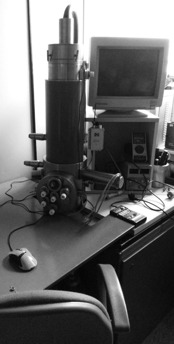
Figure 1: Modified SEM column for microwave, optical, and photochemical investigations of the living tissues.
The proposed design consists of a column in which a microscopic tube is placed, moved using the stepper motors (equipped with an opaque illuminator and an Ocean Optics fiber optic spectrofluorimeter mounted on a turret attachment) and on which an optical detection unit, a pulsed power supply, and a magnetron module with a pre-exposure camera are located. In the latter a sample is irradiated and then observed. The installation also includes an exposure control panel and a computer with software for data acquisition and processing. In the original version, the system operates at the relevant frequency of 2.45 GHz. Power and pulse modes are controlled by a panel unit, functionally similar to that of a microwave oven.
Theoretically, based on the medical physics data, it may also be appropriate to perform measurements at other frequencies (for example, 900 MHz, 930 MHz, and 1.8 GHz), at which the ROS generation and oxidative stress phenomena are recorded [21-23] (while at 1.9 GHz these data are not confirmed [24]). For this purpose, the installation provides the possibility to replace the magnetron and the exposure control panel. A noise meter was selected for this wavelength range, intended for electromagnetic noise protection measurements, since it is known that with an increase in the ROS concentrations and the DNA damage induced by them [25], electromagnetic noise can inhibit this fragmentation, leading to decreased ROS concentration [26].
The measurements are carried out in conditions protected from light (the gateway is closed at the time of exposure) in order to exclude non-thermal effects not envisaged by many authors (but also acting on the tissues) and to indicate only the response to the microwave exposure, in accordance with the work [27]. It should be noted that the authors were unable to completely standardize the microwave radiation by the wavelength, since at microwave frequencies, the magnetron operates across a range of 2.3 to 2.7 GHz and it is almost impossible to level out the parasitic modulation of the magnetron within its operational voltage range (3.5 - 4 kV). Otherwise, this installation is standardized to the levels where, according to literature, fluctuations in readings do not alter the nature of observed physiological effects. The design incorporates double shielding, ensuring complete safety for the operator. This installation can also implement fluorescence mapping techniques using specific ROS-sensitive dyes and fluorescent sensors [28].
In this case, the most oxidized areas in the sample may not directly correspond to ROS-generating locations due to the different oxidability of the macromolecular cell components, organelles, and the permeability differences for different compartments. In this regard, to objectively compare singlet oxygen generation efficiency in different sample areas, it is necessary to use not the indirect methods of ROS determination by the oxidation products, but the direct (in particular, fluorescent) methods for detecting reactive oxygen species in situ during separation, which requires the development of the control algorithms for the mechatronic subsystem of the installation corresponding to the specific exposure conditions.
In other words, in a number of experimental techniques, this installation will make it possible to map not the redox status of the cell as a final chemical outcome, but rather its lability or resistance, reflecting the physiological status, oxidability of a particular area depending on the microwave irradiation parameters. This is a qualitatively new form of functional-morphological and clinical-histochemical interpretation of in situ analysis, taking into account the form and parameters of the inductor, which were not fully described in this study [29].
Since microwaves at a wavelength of 2.45 GHz provide heating of the biological structures, and the heating of a particular structure depends on the water content, high-resolution thermography can be performed over an extended dynamic range in this installation - the so-called NIR-HDRI thermography (or bolometric measurements using enthrakometers [30]), with the co-localization analysis of the ROS and water content and thermographic characteristics mapped under the microwave irradiation. Accordingly, in the dynamic version, the technique proposed can be partially used to analyze diffusion processes and heat and mass transport. This may be useful not so much for monitoring the living cells or tissues, which are typically destroyed during such sample preparation, but for monitoring vesicular transporters of the pharmaceuticals at the preclinical stage.
It is noteworthy that the first installation shown in Figure 1 was not optimal in terms of the ratio of the chamber size and the wavelength used, that is: the working chamber as a resonator formed suboptimal configurations of standing waves with the multiple reflections from the walls, therefore producing overheating, heating gradients, and operational instability due to the insufficient chamber volume. Therefore, while formulating the priority, we cannot yet suggest the prospects for immediate introducing of this tool into the widespread biomedical practice, since its numerical optimization is required using HFSS (from ANSYS) or its analogs.
Thus, this is the first system designed to study ROS production depending on the irradiation parameters; to determine co-localization with other physicochemical parameters related to irradiation; to perform microwave heating thermography and to quantify analyte concentration; to employ fluorescent techniques using fiber optic spectrofluorimetry in order to perform direct detection of the ROS generated.
- Kochevar IE, Lambert CR, Lynch MC, Tedesco AC. Comparison of photosensitized plasma membrane damage caused by singlet oxygen and free radicals. Biochim Biophys Acta. 1996;1280(2):223-30. Available from: https://doi.org/10.1016/0005-2736(95)00297-9
- He YY, Council SE, Feng L, Bonini MG, Chignell CF. Spatial distribution of protein damage by singlet oxygen in keratinocytes. Photochem Photobiol. 2008;84(1):69-74. Available from: https://doi.org/10.1111/j.1751-1097.2007.00199.x
- Di Mascio P, Kaiser SP, Devasagayam TP, Sies H. Biological significance of active oxygen species: in vitro studies on singlet oxygen-induced DNA damage and on the singlet oxygen quenching ability of carotenoids, tocopherols and thiols. Adv Exp Med Biol. 1991;283:71-7. Available from: https://doi.org/10.1007/978-1-4684-5877-0_7
- Ray RS, Mujtaba SF, Dwivedi A, Yadav N, Verma A, Kushwaha HN, et al. Singlet oxygen mediated DNA damage induced phototoxicity by ketoprofen resulting in mitochondrial depolarization and lysosomal destabilization. Toxicology. 2013;314(2-3):229-37. Available from: https://doi.org/10.1016/j.tox.2013.10.002
- Fenoglio I, Ponti J, Alloa E, Ghiazza M, Corazzari I, Capomaccio R, et al. Singlet oxygen plays a key role in the toxicity and DNA damage caused by nanometric TiO₂ in human keratinocytes. Nanoscale. 2013;5(14):6567-76. Available from: https://doi.org/10.1039/c3nr01191g
- Rokitskaya TI, Kotova EA, Agapov II, Moisenovich MM, Antonenko YN. Unsaturated lipids protect the integral membrane peptide gramicidin A from singlet oxygen. FEBS Lett. 2014;588(9):1590-5. Available from: https://doi.org/10.1016/j.febslet.2014.02.046
- Zarębski M, Kordon M, Dobrucki JW. Photosensitized damage inflicted on plasma membranes of live cells by an extracellular generator of singlet oxygen--a linear dependence of a lethal dose on light intensity. Photochem Photobiol. 2014;90(3):709-15. Available from: https://doi.org/10.1111/php.12216
- Savin YV, Goryachev LV, Adamenkov YA, Rakhimova TV, Mankelevich YA, Popov NA, et al. Singlet oxygen production and quenching mechanisms in travelling microwave discharges. J Phys D Appl Phys. 2004;37(22):3121-8. Available from: http://dx.doi.org/10.1088/0022-3727/37/22/010
- Popović S, Rašković M, Kuo SP, Vušković L. Reactive oxygen emission from microwave discharge plasmas. J Phys Conf Ser. 2007;86:012013. Available from: http://dx.doi.org/10.1088/1742-6596/86/1/012013
- Pitz GA, Lange MA, Perram GP. Singlet oxygen kinetics in a double microwave discharge. In: High-Power Laser Ablation V. 2004;5448:1039-48. Available from: https://doi.org/10.1117/12.548707
- Yu Y, Zhang T, Zheng L, Yu J. Detection of reactive oxygen species generated by microwave electrodeless discharge lamp and application in photodegradation of H₂S. Korean J Chem Eng. 2013;30:1423-8. Available from: https://doi.org/10.1007/s11814-013-0074-z
- Lange MA, Pitz GA, Perram GP. Effect of residence time on singlet oxygen production in microwave and RF discharges. In: AIP Conf Proc. 2010;1278:482-91. Available from: https://doi.org/10.1063/1.3507137
- Özorak A, Nazıroğlu M, Çelik Ö, Yüksel M, Özçelik D, Özkaya MO, et al. Wi-Fi (2.45 GHz)- and mobile phone (900 and 1800 MHz)-induced risks on oxidative stress and elements in kidney and testis of rats during pregnancy and the development of offspring. Biol Trace Elem Res. 2013;156(1-3):221-9. Available from: https://doi.org/10.1007/s12011-013-9836-z
- Saygin M, Caliskan S, Karahan N, Koyu A, Gumral N, Uguz A. Testicular apoptosis and histopathological changes induced by a 2.45 GHz electromagnetic field. Toxicol Ind Health. 2011;27(5):455-63. Available from: https://doi.org/10.1177/0748233710389851
- Gumral N, Naziroglu M, Koyu A, Ongel K, Celik O, Saygin M, et al. Effects of selenium and L-carnitine on oxidative stress in blood of rat induced by 2.45-GHz radiation from wireless devices. Biol Trace Elem Res. 2009;132(1-3):153-63. Available from: https://doi.org/10.1007/s12011-009-8372-3
- Türker Y, Nazıroğlu M, Gümral N, Celik O, Saygın M, Cömlekçi S, et al. Selenium and L-carnitine reduce oxidative stress in the heart of rat induced by 2.45-GHz radiation from wireless devices. Biol Trace Elem Res. 2011;143(3):1640-50. Available from: https://doi.org/10.1007/s12011-011-8994-0
- Gradov OV. Experimental setups for ozonometric microscopy. Biomed Eng. 2013;46(6):260-4.
- Gradov OV. [Experimental setups for ozonometric microscopy]. Med Tekh. 2012;(6):42-7. Russian. PMID: 23304991. Available from: https://doi.org/10.1007/s10527-013-9319-8
- Yin G, Liao PH, Lo KV. An ozone/hydrogen peroxide/microwave-enhanced advanced oxidation process for sewage sludge treatment. J Environ Sci Health A Tox Hazard Subst Environ Eng. 2007;42(8):1177-81. Available from: https://doi.org/10.1080/10934520701418706
- Wong WT, Chan WI, Liao PH, Lo KV. A hydrogen peroxide/microwave advanced oxidation process for sewage sludge treatment. J Environ Sci Health A Tox Hazard Subst Environ Eng. 2006;41(11):2623-33. Available from: https://doi.org/10.1080/10934520600928086
- Zmyślony M, Politanski P, Rajkowska E, Szymczak W, Jajte J. Acute exposure to 930 MHz CW electromagnetic radiation in vitro affects reactive oxygen species level in rat lymphocytes treated by iron ions. Bioelectromagnetics. 2004;25(5):324-8. Available from: https://doi.org/10.1002/bem.10191
- Kesari KK, Kumar S, Behari J. 900-MHz microwave radiation promotes oxidation in rat brain. Electromagn Biol Med. 2011;30(4):219-34. Available from: https://doi.org/10.3109/15368378.2011.587930
- Ni S, Yu Y, Zhang Y, Wu W, Lai K, Yao K. Study of oxidative stress in human lens epithelial cells exposed to 1.8 GHz radiofrequency fields. PLoS One. 2013;8(8):e72370. Available from: https://doi.org/10.1371/journal.pone.0072370
- Brescia F, Sarti M, Massa R, Calabrese ML, Sannino A, Scarfì MR. Reactive oxygen species formation is not enhanced by exposure to UMTS 1950 MHz radiation and co-exposure to ferrous ions in Jurkat cells. Bioelectromagnetics. 2009;30(7):525-35. Available from: https://doi.org/10.1002/bem.20502
- Campisi A, Gulino M, Acquaviva R, Bellia P, Raciti G, Grasso R, et al. Reactive oxygen species levels and DNA fragmentation on astrocytes in primary culture after acute exposure to low intensity microwave electromagnetic field. Neurosci Lett. 2010;473(1):52-5. Available from: https://doi.org/10.1016/j.neulet.2010.02.018
- Yao K, Wu W, Wang K, Ni S, Ye P, Yu Y, et al. Electromagnetic noise inhibits radiofrequency radiation-induced DNA damage and reactive oxygen species increase in human lens epithelial cells. Mol Vis. 2008;14:964-9. Available from: https://pmc.ncbi.nlm.nih.gov/articles/PMC2391079/
- Marjanović AM, Pavičić I, Trošić I. Biological indicators in response to radiofrequency/microwave exposure. Arh Hig Rada Toksikol. 2012;63(3):407-16. Available from: https://doi.org/10.2478/10004-1254-63-2012-2215
- Mishina NM, Tyurin-Kuzmin PA, Markvicheva KN, Vorotnikov AV, Tkachuk VA, Laketa V, et al. Does cellular hydrogen peroxide diffuse or act locally? Antioxid Redox Signal. 2011;14(1):1-7. Available from: https://doi.org/10.1089/ars.2010.3539
- Gradov OV, Jablokov AG. Novel morphometrics-on-a-chip: CCD- or CMOS-lab-on-a-chip based on discrete converters of different physical and chemical parameters of histological samples into the optical signals with positional sensitivity for morphometry of non-optical patterns. J Biomed Technol. 2016;(2):1-29. Available from: http://dx.doi.org/10.15393/j6.art.2016.3642
- Gradov OV, Gradova MA. Microwave enthrakometric labs-on-a-chip and on-chip enthrakometric catalymetry: from non-conventional chemotronics towards microwave-assisted chemosensors. Chemosensors. 2019;7(4):48. Available from: https://doi.org/10.3390/chemosensors7040048