
More Information
Submitted: September 18, 2023 | Approved: October 10, 2025 | Published: October 13, 2025
How to cite this article: Hassan AY. Estrogen-driven Crosstalk among RUNX-2, PDLIM3, and Novel microRNAs via ERG Signaling: A Network Meta-Analysis using IPA. Arch Biotechnol Biomed. 2025; 9(1): 022-028. Available from:
https://dx.doi.org/10.29328/journal.abb.1001046.
DOI: 10.29328/journal.abb.1001046
Copyright license: © 2025 Hassan AY. This is an open access article distributed under the Creative Commons Attribution License, which permits unrestricted use, distribution, and reproduction in any medium, provided the original work is properly cited.
Keywords: Estrogen; Estrogen receptor; Estrogen signaling pathway; RUNX-2; PDLIM3; IPA tool; Regulatory microRNA

Estrogen-driven Crosstalk among RUNX-2, PDLIM3, and Novel microRNAs via ERG Signaling: A Network Meta-Analysis using IPA
Alshymaa Y Hassan*
College of Arts and Sciences, Seton Hall University, USA
*Address for Correspondence: Alshymaa Y Hassan, College of Arts and Sciences, Seton Hall University, USA, Email: [email protected]
Estrogens govern the female reproductive cycle indefinitely. Estrogens, including estrone (E1), Estradiol (E2), estriol (E3), and estetrol (E4) regulate the female life cycle from early embryonic stages and play a crucial role in development, metabolism, and cell function. Throughout evolution, estrogen has regulated reproduction by influencing the development of reproductive organs and behavior. Estrogen impacts all vertebrates, including fish, and has a role in physiological and pathological states in both genders. The RUNX-2 gene is a member of the RUNX family of transcription factors and encodes a nuclear protein with a Runt DNA-binding domain. This protein is essential for osteoblastic differentiation and skeletal morphogenesis and acts as a scaffold for nucleic acids and regulatory factors involved in skeletal gene expression. The protein can bind DNA both as a monomer or, with more affinity, as a subunit of a heterodimeric complex. In 2022, a study was conducted to characterize novel genes that are regulated by estrogen binding to its receptors (α or β). The PDLIM3 gene, with a coefficient of variation (CV) of 0.083, received the most stable CV score among other genes.
Strong correlation between estrogen binding to its receptors α or β was found, followed by expression of the PDLIM3 gene, and activation of RUNX-2 expression through the regulation of specifically miR-9 and miR-10. Also, a novel miRNA was determined, which is integrated in the activation of RUNX-2 through transcription of the PDLIM3 gene.
Estrogen, a steroid hormone playing a fundamental role in the female reproductive tract, is an essential regulator of reproductive physiology. Besides its traditional roles, estrogen possesses neuroprotective activity in preventing neurodegenerative disorders such as dementia and reducing the severity of traumatic brain injury. It is also widely utilized in Hormone Replacement Therapy (HRT) for the treatment of symptoms of menstrual irregularities and menopause [1,2].
Among all of the endogenous estrogens, 17β-estradiol (E2) is the most potent and biologically active form found in systemic circulation. E2 regulates a wide array of physiological events in diverse tissues and organs by diffusing through the plasma membrane of target cells and binding to intracellular estrogen receptors (ERs), i.e., ERα and ERβ [2,3]. These bindings trigger cascades of signals that can be divided into genomic and non-genomic mechanisms.
In genomic mechanisms, the estradiol-ER complex undergoes a hormone-binding conformational change, translocates into the nucleus, and binds to estrogen response elements (EREs) in enhancer regions, promoters, or untranslated regions of estrogen-responsive genes [4,5]. This receptor-DNA interaction controls gene transcription and subsequent protein synthesis. Otherwise, estrogen may act with membrane-bound receptors such as GPER1 or cytoplasmic receptors.
ERs in non-genomic signaling, triggering rapid activation of intracellular signaling cascades independent of genomic interaction [6,7]. Both modes of action point to the sophistication and flexibility of the hormone's function in human physiology. The complex moves to the nucleus and attaches to chromatin at ERE sequences, enhancer regions near promoters, and 30-untranslated regions of target genes (Figure 1a & 1b).
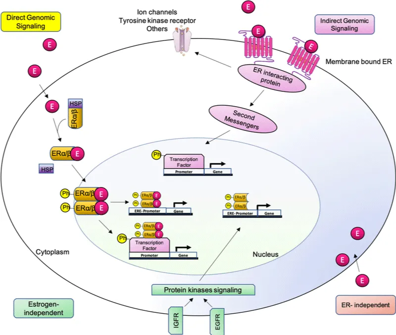
Figure 1a: Estrogen signaling pathways, including genomic and non-genomic. There are various estrogen-mediated signaling pathways. Direct genomic signaling occurs when estrogen binds to ERs. The complex dimerizes and translocates to the nucleus, causing transcriptional alterations in estrogen-responsive genes (with or without EREs). (2) Indirect genomic signaling occurs when a membrane-bound receptor modulates ion channels, second-messenger cascades, and transcription factors. (3) ER-independent: Estrogen has antioxidant properties without requiring ER activation. (4) Estrogen-independent: ligand-free genomic event; Adopted from, doi: 10.1016/bs.apcsb.2019.01.001
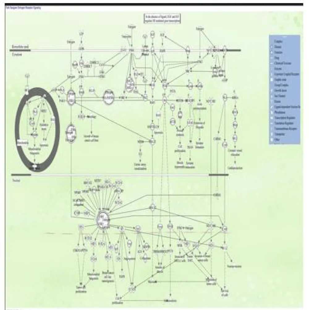
Figure 1b: Estrogen receptor signaling pathway, illustrating the key interactions and regulatory mechanisms involved. It includes elements like estrogen receptors (ERα and ERβ), heat shock proteins (HSPs), and various signaling molecules, highlighting processes such as gene expression, cell proliferation, and apoptosis. This pathway plays a crucial role in physiological functions and is particularly important in understanding conditions like breast cancer and hormone-related disorders. ERα, ERβ – Estrogen Receptor Alpha and Beta | E2 – Estradiol | HSP – Heat Shock Protein | SRC – Steroid Receptor Coactivator | PI3K – Phosphoinositide 3Kinase | AKT – Protein Kinase B | MAPK – Mitogen-Activated Protein Kinase | NF-κB – Nuclear Factor Kappa B | p53 – Tumor Protein p53 | AP-1 – Activator Protein 1 | CREB – cAMP Response Element-Binding Protein | c-Myc – Cellular Myc Protein | STAT – Signal Transducer and Activator of Transcription | EGFR – Epidermal Growth Factor Receptor | GR – Glucocorticoid Receptor | IGF-1 – Insulin-Like Growth Factor 1 | Ras – Small GTPase involved in cell signaling | JNK – c-Jun N-terminal Kinase | Fos – Proto-oncogene c-Fos | Jun – Proto-oncogene c-Jun | VEGF – Vascular Endothelial Growth Factor | Cyclin D – Cell Cycle Regulatory Protein | CDK –CyclinDependent Kinase | PTEN – Phosphatase and Tensin Homolog | GSK3β – Glycogen Synthase Kinase 3 Beta | IRS-1 – Insulin Receptor Substrate 1 | MEK – Mitogen-Activated Protein Kinase Kinase | mTOR – Mechanistic Target of Rapamycin | BCL-2 – B-Cell Lymphoma 2 | BAX – Bcl-2-Associated X Protein---designed using IPA_QIAGEN 6
The RUNX2 gene in humans encodes the transcription factor known as Runt-related transcription factor 2 (RUNX2) or core-binding factor subunit alpha-1 (CBFα1). RUNX2 is recognized as an early marker of osteogenic differentiation and plays a pivotal role in initiating osteoblast-specific extracellular matrix (ECM) synthesis by regulating the expression of critical matrix proteins such as collagen type I and osteopontin (OPN) [8]. It functions as a key transcriptional regulator of osteoblast lineage commitment.
RUNX2 encodes a nuclear-localized transcription factor containing a conserved Runt homology domain, which is essential for osteoblast differentiation and skeletal morphogenesis. It operates as a molecular scaffold for nucleic acids and transcriptional co-regulators involved in the control of skeletal gene expression [9].
Also, RUNX2 can bind DNA either as a monomer or with greater affinity when part of a heterodimeric complex. The N-terminal domain of the protein includes two potential trinucleotide repeat expansions, which, along with other mutations in the gene, have been implicated in the skeletal disorder cleidocranial dysplasia (CCD) [10].
More recently, somatic mutations in the RUNX2 gene, along with its distinct expression signatures in both healthy and neoplastic tissues, they have been highlighted for their prognostic and diagnostic value in multiple human malignancies, supporting their consideration as a cancer biomarker. Studies have demonstrated that RUNX2 contributes to the regulation of essential oncogenic processes, including tumor cell proliferation, angiogenesis, metastasis, cancer stem cell maintenance, and resistance to chemotherapy. These findings underscore the need for deeper investigation into RUNX2-mediated mechanisms as a foundation for novel therapeutic strategies [11].
The actin-associated LIM protein (ALP), product of the PDLIM3 gene, also called PDZ and LIM domain protein 3, is a structural and signaling protein found primarily in Z-discs and intercalated discs of cardiac and skeletal muscle tissue. ALP has a key role in the structural integrity of muscle as it has a role in crosslinking actin filaments via alpha-actinin-2 and is also involved in right ventricle development and functional contractility [12]. Its dysfunction has been implicated in the development of dilated cardiomyopathy (DCM), muscular dystrophy, and tumor growth, highlighting its function in biological and clinical conditions.
Despite mounting evidence for the involvement of PDLIM3 in muscle and cardiac physiology, its direct prognostic significance and immunological role within the tumor microenvironment, as in gastric cancer, remains ill-defined [13]. Unexpectedly, in 2022, PDLIM3 was reported as part of a panel of estrogen-responsive genes (ERGs).
Estrogen has a considerable effect on gene expression by its interaction with nuclear receptors. ERα and ERβ, thus either promoting or suppressing transcriptional activity. Notably, PDLIM3 has been recognized as one of the most sensitive targets under this regulatory mechanism, with a coefficient of variation (CV) of 0.083, which reflects tight regulation by estrogenic signaling [14].
Meanwhile, microRNAs (miRNAs), small non-coding RNAs approximately 19 to 25 Long nucleotides have emerged as key regulators of post-transcriptional gene expression in a variety of developmental and disease settings. Previously disregarded as genomic "noise," miRNAs are now known to inhibit gene expression, regulate cellular homeostasis, and coordinate responses in diseases from autoimmune disorders, cancer development, and viral infection [15].
In this regulatory framework, miR-9 and miR-10 are interesting because of their roles in osteogenic differentiation, whereas mechanistic pathways are still incompletely understood. Western blot studies have shown both miRNAs to influence the expression of Runt-related transcription factor- 2 (RUNX2) and the extracellular signal-regulated kinase (ERK) pathway, hypothesizing an intimate interaction between miRNA signaling and osteogenesis [16].
In addition, downregulation of miR-9 in postmitotic neurons is linked to neurodegenerative disorders, emphasizing its role in neuronal survival and maintenance [17]. miR-10 has been demonstrated to suppress T-cell proliferation, induce apoptosis, and facilitate tumor development through multiple models [17]. Interestingly, miR-9 has also been shown to promote differentiation and immunosuppressive activity of myeloid-derived suppressor cells (MDSCs) through targeting Runx1, with possible implications in immunomodulation and tumor immunity [18].
Moreover, miR-10a, a strongly conserved microRNA, has also been involved in various pathological processes, such as rheumatoid arthritis [19], juvenile dermatomyositis [20], and a range of cancers [21], underlining its therapeutic and diagnostic utility in a variety of clinical settings.
Previous functional studies have also corroborated the idea that miR-10a-3p actively suppresses the production of Inhibitors of differentiation (ID) genes ID3, boosting the activity of the ossification core factor RUNX2 [22]. Another microRNA, which was highlighted, is microRNA6769B (Mammalian), which was discovered to have an indirect regulatory influence on the expression of the RUNX-2 gene via miR1896 (and other miRNAs w/seed GGUGGGU) (Mammalian) activation, leading to upregulation of downstream signaling pathways.
The purpose of our research is to shed light on current mechanistic findings and the modulatory role of estrogen via both direct and indirect effects on the signaling pathways that regulate RUNX-2 expression. Depending on the data analyzed, our primary goal is to link the expression and regulation of microRNA9, microRNA10, miR-1896, and microRNA6769B to RUNX-2 expression by modulating the new estrogen receptor gene, ERG-PDLIM3. QIAGEN's bioinformatics tool, Ingenuity Pathway Analysis (IPA), was used to design molecular networks and analyze their biological roles. The molecular networks were compared to QIAGEN Knowledge Base (QKB) findings using canonical and signaling pathway analysis, as well as other statistical approaches.
Ingenuity pathway analysis software
IPA, a bioinformatics software tool for data mining, uses canonical pathways and gene regulatory networks from the literature to help interpret and analyze various biological pathways. A variety of techniques were used to create pathways depicting the molecular networks connected with estrogen, RUNX-2, different miRNAs, and their intermediary molecules to evaluate functional hypotheses. The bioinformatics tool utilized data from the QIAGEN Knowledge Base (QKB) between February 5th, 2024 and June 14th, 2025 [23-26]. The workflow utilized from QIAGEN’s Ingenuity Pathway Analysis (IPA) bioinformatics software for data mining is illustrated in Figure 2.
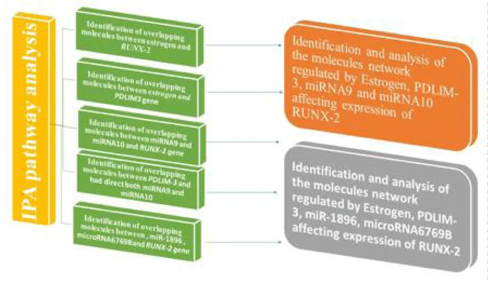
Figure 2: he data mining workflow was based on QIAGEN's Ingenuity Pathway Analysis (IPA) bioinformatics tools. The "Grow","Connect", "Pathway Explorer", and "Molecule Activity Predictor" (MAP) tools from the "My Pathway" feature were used to develop biological networks that showed the connectivity between distinct nodes. Furthermore, the "Core Analysis: Expression Analysis" tool was utilized to compare the molecules within the produced molecular route to canonical pathways recorded within QIAGEN's knowledge base (QKB).
Molecular pathway analysis of molecules mediating the relationship between estrogen and PDLIM3
The "MAP" program was used to generate a connectivity map depicting the interaction between the 10 molecules involved with estrogen's direct influence and their relationship with PDLIM3. Our findings show that estrogen is associated with intermediates such as Proliferating Cellular Nuclear Antigen (PCNA), Cyclin-Dependent Kinase 4 (CDK4), Luteinizing Hormone (LH), Mitogen-Activated Protein Kinase-1 (MAPK-1), Estrogen Receptor 1 and 2 (ESR 1 & 2), and Follicle-Stimulating Hormone (FSH), all of which are linked to the PDLIM3 gene.
Molecular pathway analysis of molecules mediating the relationship between estrogen and affect expression of RUNX-2
The "MAP" program and QKB can identify 75 estrogen-controlled pathways that regulate RUNX-2 expression. These molecules included biological medicines, the canonical pathway, complexes, cytokines, enzymes, G-protein coupled receptors, kinases, nuclear receptors, peptidases, phosphatases, transcription and translation regulators, transmembrane receptors, and transporters.
Molecular pathway analysis of molecules mediating the relationship between miRNA9 and directly affecting the expression of the RUNX-2 gene
The "MAP" program and QKB identified 8 pathways controlled by miRNA9 that modulate RUNX-2 expression. These molecules comprised G-protein coupled receptors, kinases, nuclear receptors, transcription and translation regulators, transmembrane receptors, and transporters.
Molecular pathway analysis of molecules mediating the relationship between miRNA10 and directly affecting the expression of the RUNX-2 gene
The "MAP" program and QKB identified 34 pathways controlled by miRNA10 that modulate RUNX-2 expression. These molecules comprise G-protein coupled receptors, kinases, nuclear receptors, transcription and translation regulators, transmembrane receptors, and transporters.
Molecular pathway analysis of molecules mediating the relationship between PDLIM-3 and had direct effects on the expression of miRNA9
The "MAP" program and QKB identified 49 pathways directly regulated by PDLIM-3 that modulate miRNA9 expression. These molecules comprise G-protein coupled receptors, kinases, nuclear receptors, transcription and translation regulators, transmembrane receptors, and transporters.
Molecular pathway analysis of molecules mediating the relationship between PDLIM-3 and had direct effects on the expression of miRNA-10
The "MAP" program and QKB identified 98 pathways directly regulated by PDLIM-3 that modulate miRNA9 expression. These molecules comprised G-protein coupled receptors, kinases, nuclear receptors, transcription and translation regulators, transmembrane receptors, and transporters.
Identification and analysis of the molecular network regulated by Estrogen, PDLIM-3, miRNA9, and miRNA10 affecting the expression of RUNX-2
The "MAP" program and QKB identified around 39 molecules involved directly or indirectly in the expression of RUNX-2, regulated by estrogen, miRNA9, and miRNA10, and PDLIM-3 interaction. The full classification of those molecules, including the gene name, its location, and its family.
Identification and analysis of the molecular network regulated by Estrogen, PDLIM-3, miR-1896, microRNA6769B, affecting the expression of RUNX-2
The "MAP" program and QKB identified around 45 molecules involved directly or indirectly in the expression of RUNX-2, regulated by estrogen, microRNA-1896, microRNA-6769 B, and PDLIM-3. The full classification of those molecules, including the gene name, its location, and its family.
This study elucidates a novel, multilayered regulatory axis in which estrogen activates RUNX2 expression through both direct genomic signaling and epigenetic modulation involving PDLIM3 and specific microRNAs. The identification of PDLIM3 as an intermediary, along with the integration of miR-9, miR-10, and the newly implicated miR6769b, provides fresh insight into estrogen-responsive osteogenesis and opens potential avenues for targeted therapeutic strategies.
The pathway identified can be summarized as:
Estrogen → ERα/β ERα/β → PDLIM3 ↑ → miR-9/miR-10/miR-6769b ↑→RUNX-2 ↑
Classical estrogen receptor signaling and PDLIM3 activation
Estrogen binding to ERα and ERβ activates classical genomic signaling, whereby the ligand-receptor complex translocates to the nucleus and binds estrogen response elements (EREs) in the regulatory regions of target genes [27]. Our analysis revealed a strong association between estrogen signaling and PDLIM3 expression. Although PDLIM3 has previously been associated primarily with muscle function [28], this is the first report linking it to estrogen-mediated osteogenic pathways and RUNX2 regulation. Its identification as a novel intermediary in this axis is supported by its low coefficient of variation (CV = 0.083), indicating tight estrogenic regulation [29].
PDLIM3-mediated regulation of miR-9 and miR-10
Downstream of PDLIM3, our IPA-based analysis identified significant associations with miR-9 and miR-10, two miRNAs implicated in bone formation, neurodevelopment, and immune regulation [30,31]. Both miRNAs have been shown to regulate RUNX2 directly or via ERK signaling, and their increased expression in response to estrogen-PDLIM3 signaling suggests a crucial post-transcriptional modulatory role in osteogenesis [32].
Novel discovery of miR-6769b in osteogenic regulation
An especially noteworthy finding is the identification of miR-6769b as a novel epigenetic player within this regulatory framework. While limited prior data exist linking miR-6769b to bone biology, emerging literature suggests its involvement in exosome-mediated bone remodeling and cell proliferation control [33,34]. Our network data suggest that miR-6769b is induced downstream of PDLIM3, representing a new layer of epigenetic regulation for RUNX2.
Integrated signaling cascade
Our findings support a multi-step model for estrogen-driven RUNX2 activation:
Estrogen → ERα/β → PDLIM3 ↑ → miR-9/miR-10/miR-6769b ↑ → RUNX2 ↑
This pathway expands upon the classical model of estrogen action by incorporating miRNA-mediated epigenetic regulation and identifying new targets such as PDLIM3 and miR6769b that can serve as biomarkers or therapeutic entry points in skeletal disorders [27,29].
Implications and future directions
Biological Significance: This model enriches our understanding of estrogenic regulation of skeletal development and uncovers new candidate targets for bone regeneration and disease treatment.
Experimental validation: Further experimental validation using ChIP-qPCR (to confirm ER binding to PDLIM3), gene knockdown (for PDLIM3 and miR-6769b), and miRNA modulation studies is critical to establish functional causality.
In this study, pathway relationships between estrogen, PDLIM3, microRNAs (miR-9, miR-10, and miR-6769b), and RUNX2 were primarily modeled using QIAGEN's Ingenuity Pathway Analysis (IPA) tools, in conjunction with curated biological insights from literature. While z-score-based predictions are commonly employed to infer activation or inhibition states in large-scale differential expression studies, they were not the focus of this analysis. Instead, we aimed to systematically map molecular interactions and delineate hierarchical regulatory networks rather than statistically quantify expression changes.
Consequently, the findings are presented descriptively, highlighting directionality and mechanistic connectivity as supported by canonical pathway data and peer-reviewed evidence. This approach has uncovered a novel, multilayered regulatory cascade whereby estrogen signaling through ERα/β leads to upregulation of PDLIM3, which in turn enhances the expression of key microRNAs, including miR-9, miR-10, and the newly implicated miR-6769b, ultimately driving the transcriptional activation of RUNX2.
These insights reveal new mediators within the estrogen–RUNX2 axis and offer promising implications for future research into skeletal development, osteogenic differentiation, and hormone-responsive bone pathologies such as osteoporosis and fracture repair.
I would like to express my deepest gratitude to Professor Sulie Chang and the Institute of Neuroimmune Pharmacology (INIP) at Seton Hall University for their continuous support, guidance, and mentorship throughout my graduate studies. Their invaluable contributions and encouragement have played a significant role in the development of this work and my academic growth.
- Huether SE, McCance KL. Understanding pathophysiology. 7th ed. St. Louis (MO): Mosby; 2020. Available from: https://shop.elsevier.com/books/understanding-pathophysiology/huether/978-0-323-63908-8
- Delgado BJ, Patel P, Lopez-Ojeda W. Estrogen(Archived). 2025 Aug 9. In: StatPearls [Internet]. Treasure Island (FL): StatPearls Publishing; 2025. Available from: https://pubmed.ncbi.nlm.nih.gov/30855848/
- Sier JH, Thumser AE, Plant NJ. Linking physiologically-based pharmacokinetic and genome-scale metabolic networks to understand estradiol biology. BMC Syst Biol. 2017;11(1):141. Available from: https://doi.org/10.1186/s12918-017-0520-3
- Shaban NZ, Talaat IM, Elrashidy FH, Hegazy AY, Sultan AS. Therapeutic role of Punica granatum (pomegranate) seed oil extract on bone turnover and resorption induced in ovariectomized rats. J Nutr Health Aging. 2017;21(10):1299-306. Available from: https://doi.org/10.1007/s12603-017-0884-5
- Thompson PA, Ambrosone C. Molecular epidemiology of genetic polymorphisms in estrogen metabolizing enzymes in human breast cancer. JNCI Monogr. 2000;2000(27):125-34. Available from: https://doi.org/10.1093/oxfordjournals.jncimonographs.a024235
- Fuentes N, Silveyra P. Estrogen receptor signaling mechanisms. Adv Protein Chem Struct Biol. 2019;116:135-70. Available from: https://doi.org/10.1016/bs.apcsb.2019.01.001
- Le Dily F, Beato M. Signaling by steroid hormones in the 3D nuclear space. Int J Mol Sci. 2018;19(2):306. Available from: https://doi.org/10.3390/ijms19020306
- Komori T. Runx2, an inducer of osteoblast and chondrocyte differentiation. Histochem Cell Biol. 2019;151(1):1–11.
- Lucero MJ, Vega OA, Osorio MM, Tapia JC, Antonelli M, Stein GS, Galindo MA. The cancer-related transcription factor Runx2 modulates cell proliferation in human osteosarcoma cell lines. J Cell Physiol. 2013;228(4):714–723. Available from: https://doi.org/10.1002/jcp.24218
- Lin TC. RUNX2 and cancer. Int J Mol Sci. 2023;24(8):7001. Available from: https://doi.org/10.3390/ijms24087001
- Hu X, Chen M, Ruan Q, Shi C, Pan J, Luo L. Comprehensive analysis of PDLIM3 expression profile, prognostic value, and correlations with immune infiltrates in gastric cancer. J Immunol Res. 2022;2022:2039447. Available from: https://www.hindawi.com/journals/jir/2022/2039447/
- Nishi K, Fu W, Kiyama R. Novel estrogen-responsive genes (ERGs) for the evaluation of estrogenic activity. PLoS One. 2022;17(8):e0272553. Available from: https://journals.plos.org/plosone/article?id=10.1371/journal.pone.0273164
- Ranganathan K, Sivasankar V. MicroRNAs – biology and clinical applications. J Oral Maxillofac Pathol. 2014;18(2):229–234.
- Luo H, Gao H, Liu F, Qiu B. Regulation of Runx2 by microRNA-9 and microRNA-10 modulates the osteogenic differentiation of mesenchymal stem cells. Int J Mol Med. 2017;39(5):1046–1052. Available from: https://www.spandidos-publications.com/10.3892/ijmm.2017.2918
- Jiang M, Zhang W, Zhang R, Liu P, Ye Y, Yu W, et al. Cancer exosome-derived miR-9 and miR-181a promote the development of early-stage MDSCs via interfering with SOCS3 and PIAS3, respectively, in breast cancer. Oncogene. 2020;39(24):4681–4694. Available from: https://www.nature.com/articles/s41388-020-1322-4
- Tian J, Rui K, Tang X, Ma J, Wang Y, Tian X, et al. MicroRNA-9 regulates the differentiation and function of myeloid-derived suppressor cells via targeting Runx1. J Immunol. 2015;195(3):1301–1311. Available from: https://www.jimmunol.org/content/195/3/1301
- Jiang M, Zhang W, Zhang R, Liu P, Ye Y, Yu W, et al. Cancer exosome-derived miR-9 and miR-181a promote the development of early-stage MDSCs via interfering with SOCS3 and PIAS3, respectively, in breast cancer. Oncogene. 2020;39(24):4681–4694. Available from: https://www.nature.com/articles/s41388-020-1322-4
- Mu N, Gu J, Huang T, Zhang C, Shu Z, Li M, et al. A novel NFκB/YY1/microRNA-10a regulatory circuit in fibroblast-like synoviocytes regulates inflammation in rheumatoid arthritis. Sci Rep. 2016;6:20059. Available from: https://www.nature.com/articles/srep20059
- Xu D, Huang CC, Kachaochana A, Morgan GA, Bonaldo MF, Soares MB, et al. MicroRNA-10a regulation of proinflammatory mediators: an important component of untreated juvenile dermatomyositis. J Rheumatol. 2016;43(1):161–168. Available from: https://www.jrheum.org/content/43/1/161
- Stadthagen G, Tehler D, Høyland-Kroghsbo NM, Wen J, Krogh A, Jensen KT, et al. Loss of miR-10a activates Lpo and collaborates with activated Wnt signaling in inducing intestinal neoplasia in female mice. PLoS Genet. 2013;9(10):e1003913. Available from: https://journals.plos.org/plosgenetics/article?id=10.1371/journal.pgen.1003913
- Xu C, Zhang H, Gu W, Wu H, Chen Y, Zhou W, et al. The microRNA-10a/ID3/RUNX2 axis modulates the development of ossification of the posterior longitudinal ligament. Sci Rep. 2018;8(1):9223. Available from: https://www.nature.com/articles/s41598-018-27514-x
- Bishir M, Rengifo T, Huang W, Kim RJ, Chidambaram SB, Chang SL. Network meta-analysis on alcohol-mediated modulation of Alzheimer’s disease in the diseases of inflammation, including COVID-19. NeuroImmune Pharmacol Ther. 2023;2(3):267–281.
- Rengifo T, Bishir M, Huang W, Snyder M, Chang SL. Network meta-analysis of the molecular mechanisms and signaling pathways underlying alcohol-induced thymic atrophy. Alcohol Clin Exp Res. 2024;48(5):795–809. Available from: https://doi.org/10.1111/acer.15292
- Zhang J, Bishir M, Barbhuiya S, Chang SL. Meta-analysis of the mechanisms underlying COVID-19 modulation of Parkinson’s disease. Int J Mol Sci. 2023;24(17):13554. Available from: https://doi.org/10.3390/ijms241713554
- Otto F, Thornell AP, Crompton T, Denzel A, Gilmour KC, Rosewell IR, et al. Cbfa1, a candidate gene for cleidocranial dysplasia syndrome, is essential for osteoblast differentiation and bone development. Cell. 1997;89(5):765–771. Available from: https://doi.org/10.1016/s0092-8674(00)80259-7
- Krcmery J, Camarata T, Kulisz A, Simon HG. Loss of the cytoskeletal protein α-actinin-2 leads to dilated cardiomyopathy and skeletal muscle dysfunction. Proc Natl Acad Sci U S A. 2010;107(6):2345–2350.
- Komori T. Runx2, an inducer of osteoblast and chondrocyte differentiation. Histochem Cell Biol. 2019;151(1):1–11. Available from: https://doi.org/10.1007/s00418-018-1640-6
- Zhou L, Song Z, Hu J, Gao M, Bao J, Zhang G, et al. MiR-9 promotes osteoblast differentiation and suppresses osteoclastogenesis via targeting DKK1. Gene. 2015;564(2):137–144.
- Wu D, Huang Y, Kang J, Li K, Bi X, Zhang T, et al. MiR-10a promotes osteogenesis and suppresses adipogenesis in human mesenchymal stem cells. Aging Cell. 2014;13(2):333–341.
- Sun G, Ye P, Murai K, Lang MF, Li S, Zhang H, et al. miR-9 regulates neural stem cell proliferation and differentiation by targeting TLX and REST. Dev Cell. 2011;20(6):897–909.
- Wang H, Peng W, Ouyang X, Li W, Dai Y. miR-10a regulates T-cell activation and promotes immunosuppression in ovarian cancer. Cell Death Dis. 2015;6(7):e1909.
- Liu Y, Zhang Q, Chen L, Wang X, Li J, Zhao H, et al. miR-6769b-5p targets CCND1 to regulate proliferation in cadmium-exposed placental trophoblasts. Sci Total Environ. 2022;825:153927.
- NET Research Team. Exosome-mediated regulation of bone remodeling by miRNAs, including miR-6769b. 2019.
- Hassan A. Network meta-analysis of estrogen's direct and indirect influence on signaling pathways controlling RUNX-2 through regulation of novel ERG gene. 2025. Available from: https://www.researchgate.net/publication/391160429_Network_meta-analysis_of_estrogen%27s_direct_and_indirect_influence_on_signaling_pathways_controlling_RUNX-2through_regulation_of_novel_ERG_gene