Figure 6b
OPEN and CLOSED state of SPIKE SARS-COV-2: relationship with some integrin binding. A biological molecular approach to better understand the coagulant effect
Luisetto M*, Khaled Edbey, Mashori GR, Yesvi AR and Latyschev OY
Published: 02 July, 2021 | Volume 5 - Issue 1 | Pages: 049-056
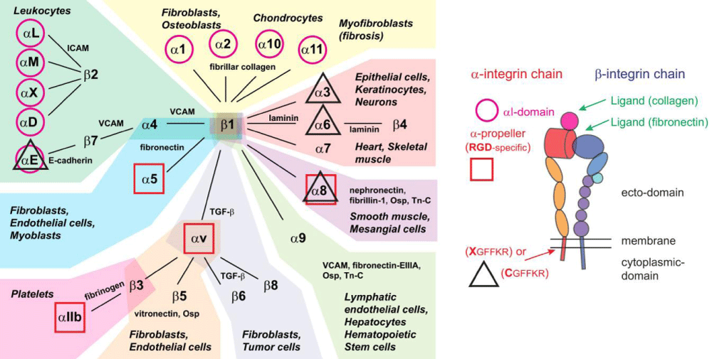
Figure 6b:
6b: Integrins, their ligands, and cellular distribution. According Hynes: integrins are organized in 24 different hetero-dimers, indicated by the link between α- and β-subunits in the figure. Integrins can be classified by structural -features, their ligands, and their tissue and cellular- expression. Based on these criteria, we grouped integrins into 9 classes indicated by the color of their background. Typical cell types expressing the respective integrins are mentioned, like common ligands for these integrins. We also highlighted integrins with an α-I domain (I; purple circle), those binding to RGD- ligands (RGD; red square), and those with changes to the conserved GFFKR sequence in the membrane-proximal part of the α-subunit (black triangle indicates integrins with sequence deviating from CGFFKR). The integrin- cartoon in the bottom part of the figure gives an overview about the integrin -structure and is reused in the following figures. It also indicates the location of the αI domain and the GFFKR sequence in the respective integrins. Please note that α-I domain integrins bind ligands (collagen) via this I domain, while other integrins bind ligands (fibronectin) in binding -pockets formed by both α- and β-subunits. Qiao B, de la Cruz MO. Enhanced Binding of SARS-CoV-2 Spike Protein to Receptor by Distal Polybasic Cleavage Sites. ACS Nano. 2020; 14: 10616–10623. https://doi.org/10.1021/acsnano.0c04798
Read Full Article HTML DOI: 10.29328/journal.abb.1001028 Cite this Article Read Full Article PDF
More Images
Similar Articles
-
OPEN and CLOSED state of SPIKE SARS-COV-2: relationship with some integrin binding. A biological molecular approach to better understand the coagulant effectLuisetto M*,Khaled Edbey,Mashori GR,Yesvi AR,Latyschev OY. OPEN and CLOSED state of SPIKE SARS-COV-2: relationship with some integrin binding. A biological molecular approach to better understand the coagulant effect . . 2021 doi: 10.29328/journal.abb.1001028; 5: 049-056
Recently Viewed
-
Evaluation of the Bone Marrow Aspirate Examination Practice at the University Hospital Andrainjato Fianarantsoa, MadagascarBronislaw Tchestérico Dodoson, Jocia Fenomanana, Manevarivo Eddy Ramaminiaina, Stéphania Niry Manantsoa, Zely Arivelo Randriamanantany, Andriamiadana Luc Rakotovao, Aimée Olivat Rakoto Alson. Evaluation of the Bone Marrow Aspirate Examination Practice at the University Hospital Andrainjato Fianarantsoa, Madagascar. J Hematol Clin Res. 2023: doi: 10.29328/journal.jhcr.1001023; 7: 015-020
-
A Short communication on Pichia pastoris vs. E. coli: Efficient expression systemViswanath Vittaladevaram*. A Short communication on Pichia pastoris vs. E. coli: Efficient expression system. Ann Proteom Bioinform. 2021: doi: 10.29328/journal.apb.1001016; 5: 049-050
-
In silico disrupting quorum sensing of porphyromonas gingivalis via essential oils and coumarin derivativesHaitham Ahmed Al-Madhagi*,Zaher Samman Tahan. In silico disrupting quorum sensing of porphyromonas gingivalis via essential oils and coumarin derivatives. Ann Proteom Bioinform. 2022: doi: 10.29328/journal.apb.1001017; 6: 001-005
-
Three-phase partitioning for isolating peroxidase from lemon peelsHaitham Ahmed Al-Madhagi*,Valantina Yazbik,Wassim Abdelwahed. Three-phase partitioning for isolating peroxidase from lemon peels. Ann Proteom Bioinform. 2023: doi: 10.29328/journal.apb.1001018; 7: 001-005
-
A brief evaluation of the concepts of omics, genomics, proteomics and metabolomics in terms of sperm cellAhmet Yörü, Belkıs Kamer, Serkan Ali Akarsu, Ali Doğan Ömür*. A brief evaluation of the concepts of omics, genomics, proteomics and metabolomics in terms of sperm cell. Ann Proteom Bioinform. 2023: doi: 10.29328/journal.apb.1001019; 7: 006-009
Most Viewed
-
Feasibility study of magnetic sensing for detecting single-neuron action potentialsDenis Tonini,Kai Wu,Renata Saha,Jian-Ping Wang*. Feasibility study of magnetic sensing for detecting single-neuron action potentials. Ann Biomed Sci Eng. 2022 doi: 10.29328/journal.abse.1001018; 6: 019-029
-
Evaluation of In vitro and Ex vivo Models for Studying the Effectiveness of Vaginal Drug Systems in Controlling Microbe Infections: A Systematic ReviewMohammad Hossein Karami*, Majid Abdouss*, Mandana Karami. Evaluation of In vitro and Ex vivo Models for Studying the Effectiveness of Vaginal Drug Systems in Controlling Microbe Infections: A Systematic Review. Clin J Obstet Gynecol. 2023 doi: 10.29328/journal.cjog.1001151; 6: 201-215
-
Prospective Coronavirus Liver Effects: Available KnowledgeAvishek Mandal*. Prospective Coronavirus Liver Effects: Available Knowledge. Ann Clin Gastroenterol Hepatol. 2023 doi: 10.29328/journal.acgh.1001039; 7: 001-010
-
Causal Link between Human Blood Metabolites and Asthma: An Investigation Using Mendelian RandomizationYong-Qing Zhu, Xiao-Yan Meng, Jing-Hua Yang*. Causal Link between Human Blood Metabolites and Asthma: An Investigation Using Mendelian Randomization. Arch Asthma Allergy Immunol. 2023 doi: 10.29328/journal.aaai.1001032; 7: 012-022
-
An algorithm to safely manage oral food challenge in an office-based setting for children with multiple food allergiesNathalie Cottel,Aïcha Dieme,Véronique Orcel,Yannick Chantran,Mélisande Bourgoin-Heck,Jocelyne Just. An algorithm to safely manage oral food challenge in an office-based setting for children with multiple food allergies. Arch Asthma Allergy Immunol. 2021 doi: 10.29328/journal.aaai.1001027; 5: 030-037

HSPI: We're glad you're here. Please click "create a new Query" if you are a new visitor to our website and need further information from us.
If you are already a member of our network and need to keep track of any developments regarding a question you have already submitted, click "take me to my Query."


